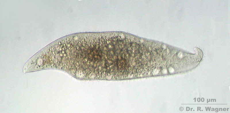
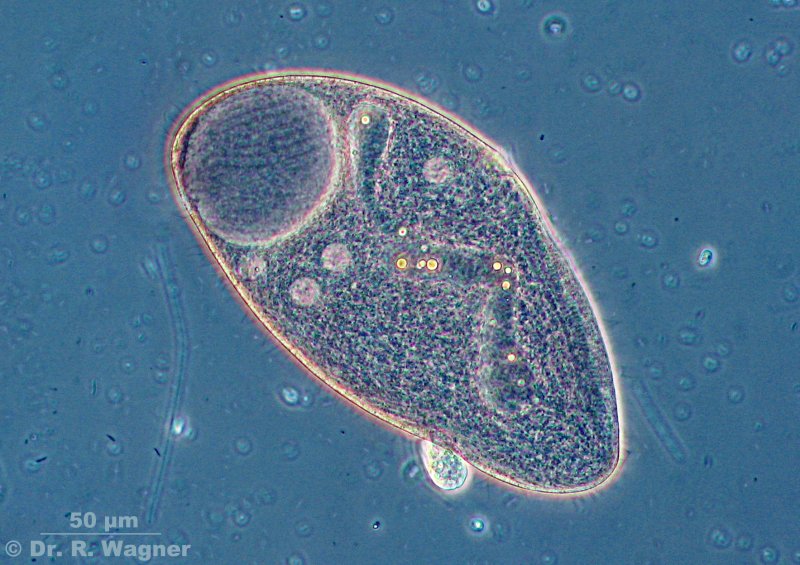
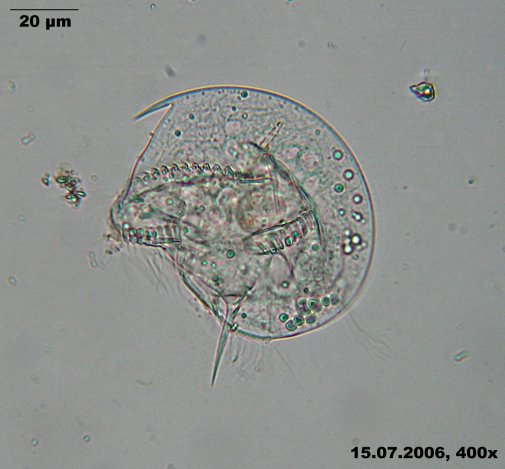
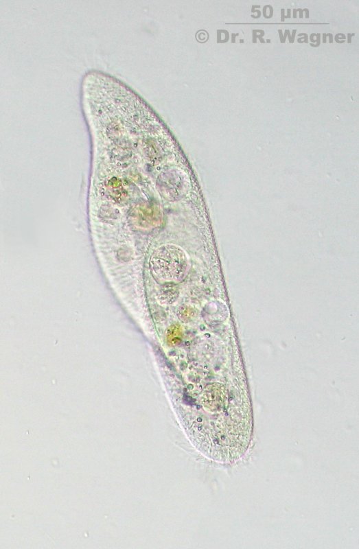
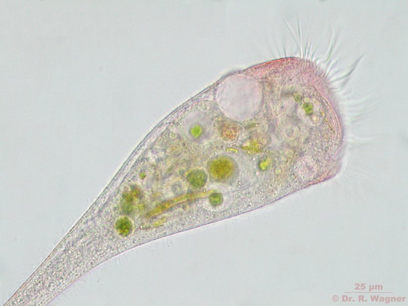
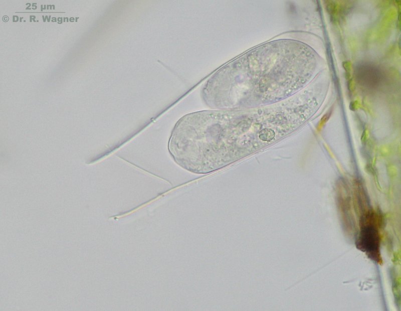
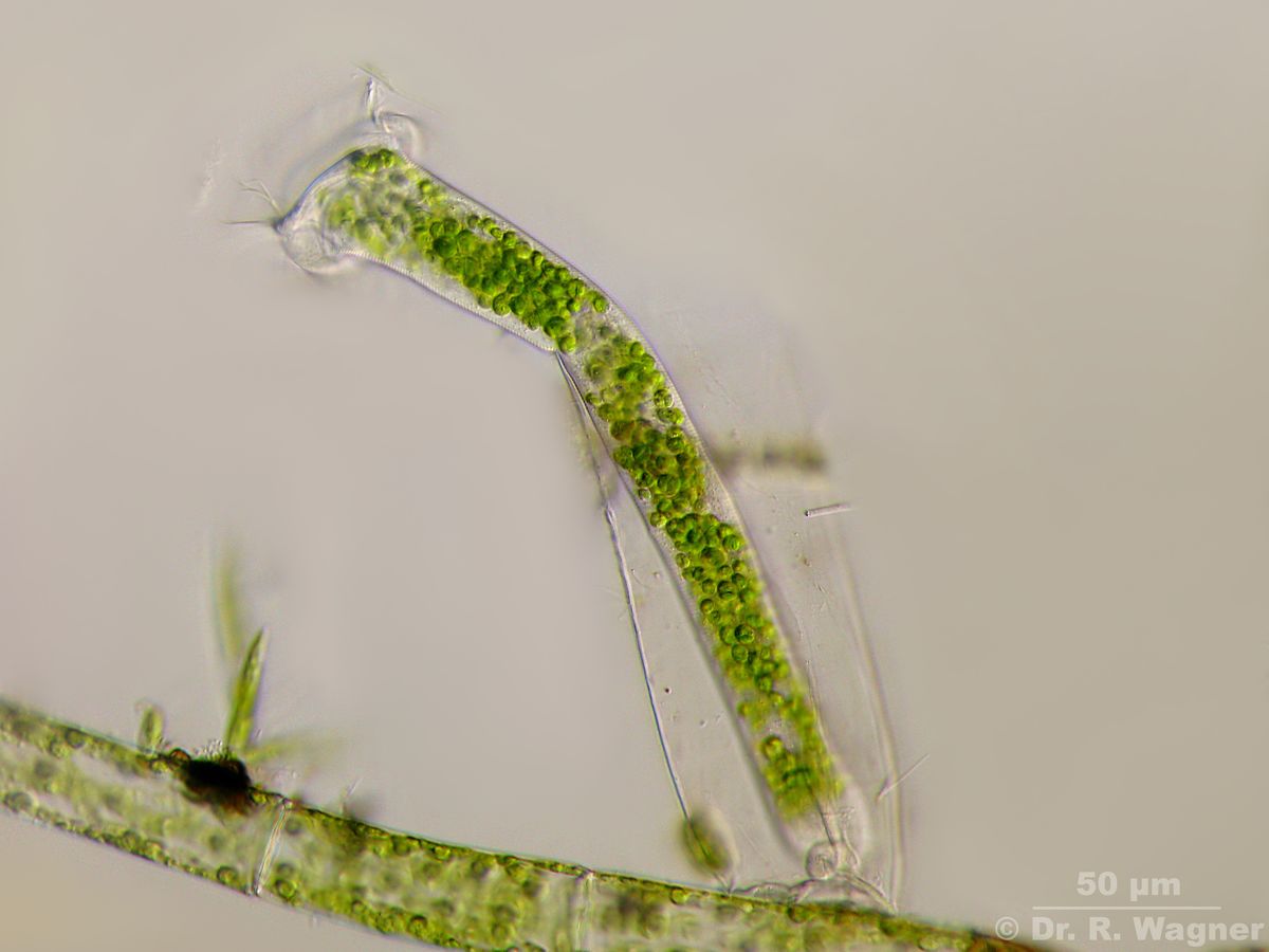
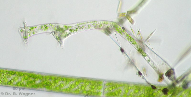
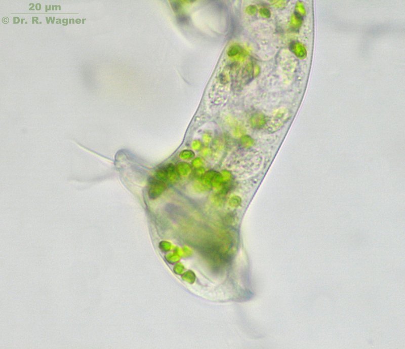
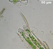
Amphileptus pleurosigma Garden pond, November 2007
Ventral and dorsal a row of vacuoles, eachMicroscopy, photomicrograph, Microfoto
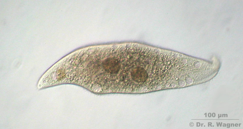
Amphileptus pleurosigma Garden pond, November 2007
In the middle 2 macronucleii, to the reight the thorn-shaped extrusoms become visible.
They are evenly distributed over the whole plasma.Microscopy, photomicrograph, Microfoto
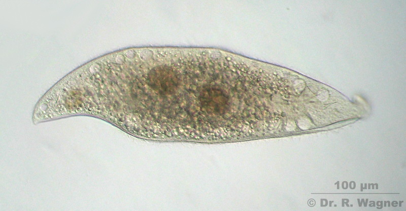
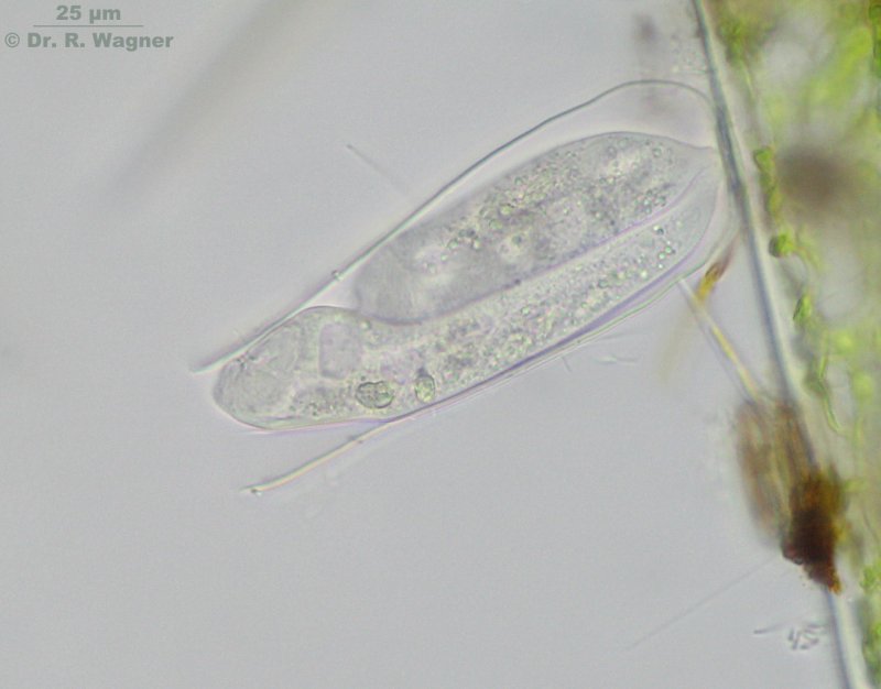
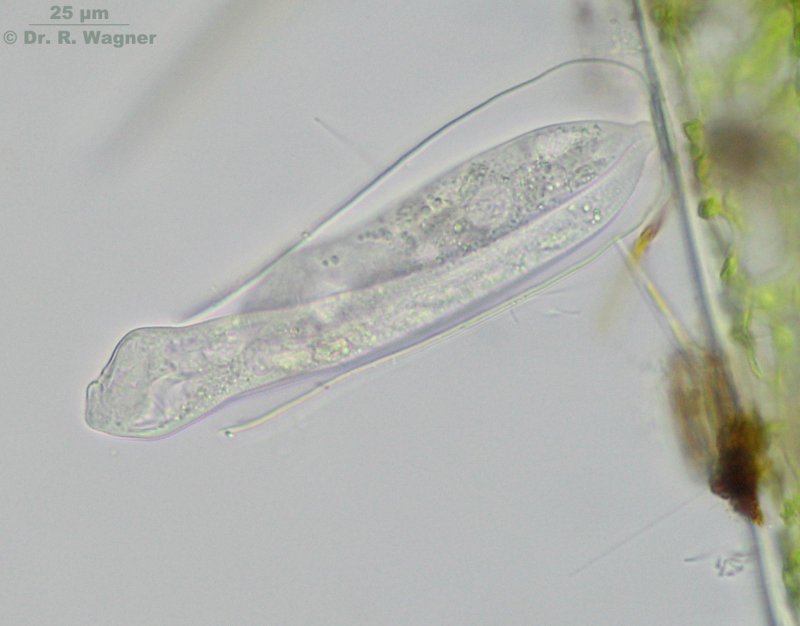
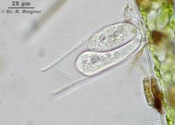
Amphileptus pleurosigma Garden pond, November 2007
Microscopy, photomicrograph, Microfoto
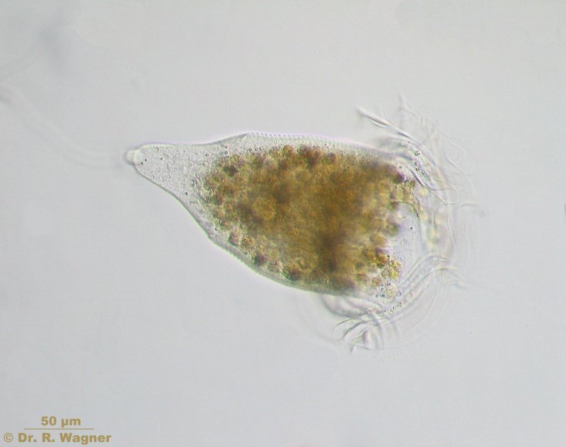
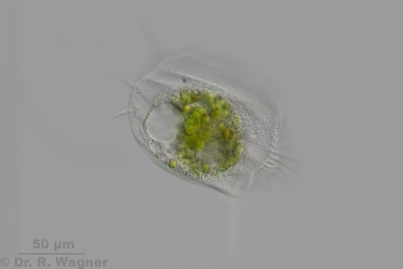
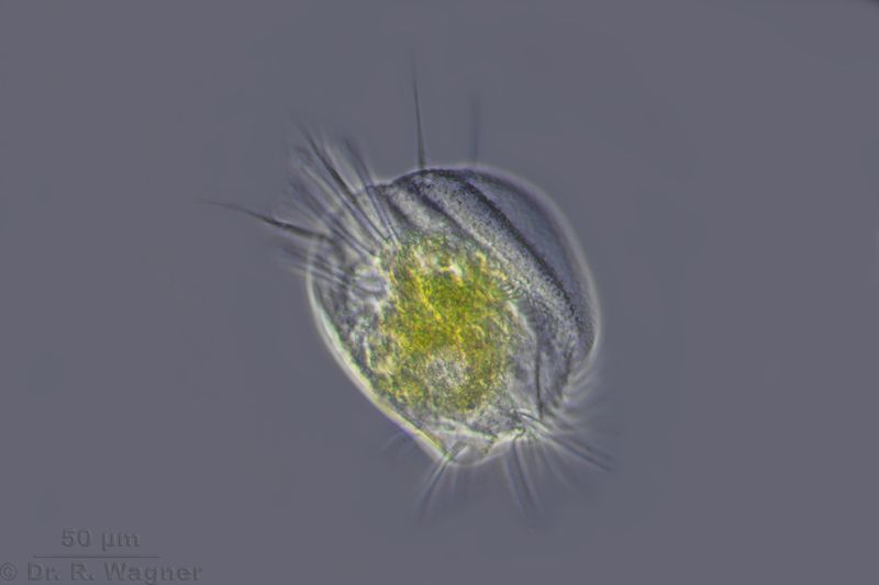
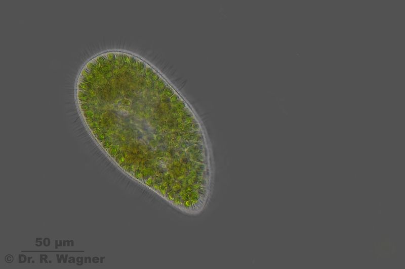
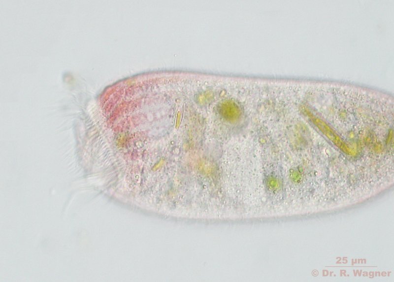
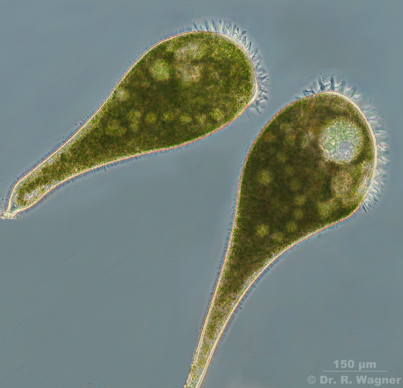
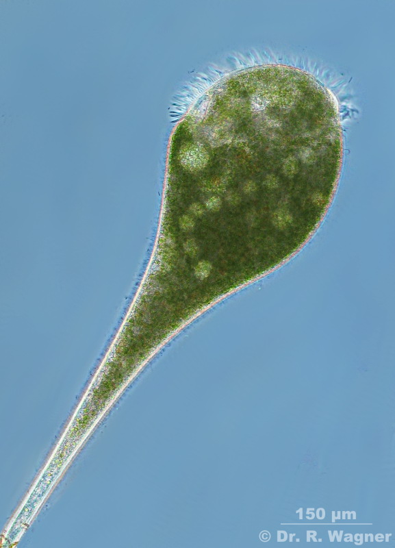
Blepharisma spec. Culture-material, June 2007Microscopy, photomicrograph, Microfoto
Blepharisma spec. Culture-material, June 2007Microscopy, photomicrograph, Microfoto
Blepharisma spec. Culture-material, June 2007Microscopy, photomicrograph, Microfoto
Phase contrast
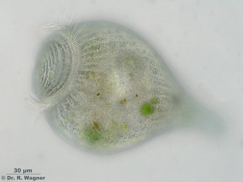
campanella umbellaria garden pond, February 2007Microscopy, photomicrograph, Microfoto
normal state
campanella umbellaria garden pond, February 2007Microscopy, photomicrograph, Microfoto
startle response
campanella umbellaria garden pond, May 2003Microscopy, photomicrograph, Microfoto
this video (AVI-format, 1.1 MB)
campanella umbellaria garten pond, March 2004Microscopy, photomicrograph, Microfoto
campanella umbellaria garden pond, January 2005
this video (AVI-(DIVX)-format, 1.7 MB) shows a swarmer.Microscopy, photomicrograph, Microfoto
cothurnia annulata garden pond, July 2005Microscopy, photomicrograph, Microfoto
cothurnia annulata garden pond, July 2005
video (2,4 MB, divx-format)Microscopy, photomicrograph, Microfoto
discomorphella pectinata garden pond, July 2006Microscopy, photomicrograph, Microfoto
2 pictures stacked with CombineZ5
Euplotes spec. heathland pond June 2015
Microscopy, photomicrograph, Microfoto
Euplotes spec. heathland pond June 2015
Microscopy, photomicrograph, Microfoto
Phasecontrast
euplotes spec. national horticultural show Duesseldorf, May 2006
Microscopy, photomicrograph, Microfoto
Euplotes patella Botanical garden Uni Duesseldorf, March 2008Microscopy, photomicrograph, Microfoto
Euplotes patella in most cases lives without some symbionts.
Only the individuals that live in sapropel do often show symbiontic green algae
Euplotes patella Botanical garden Uni Duesseldorf, March 2008Microscopy, photomicrograph, Microfoto
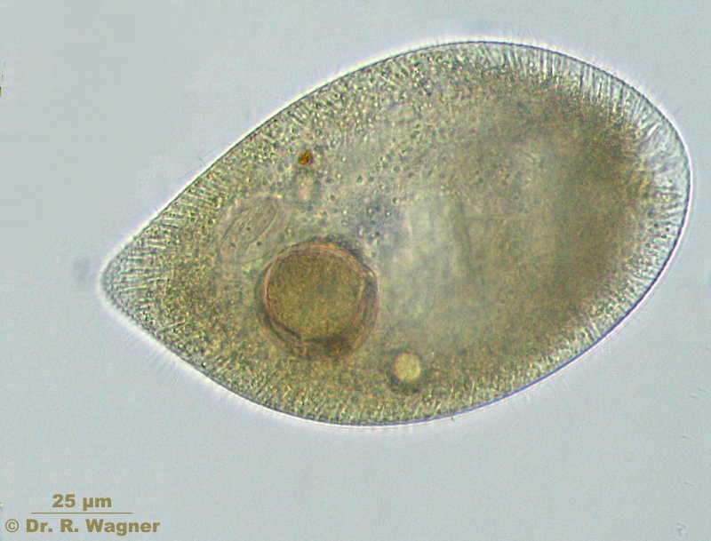
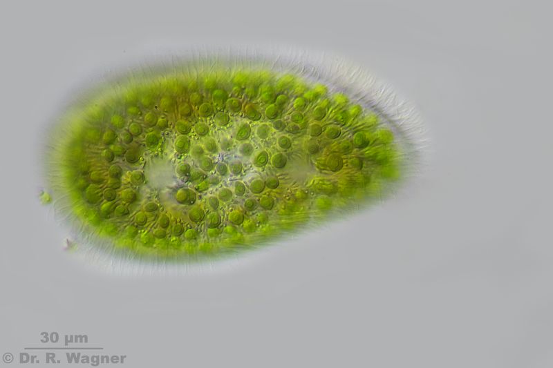
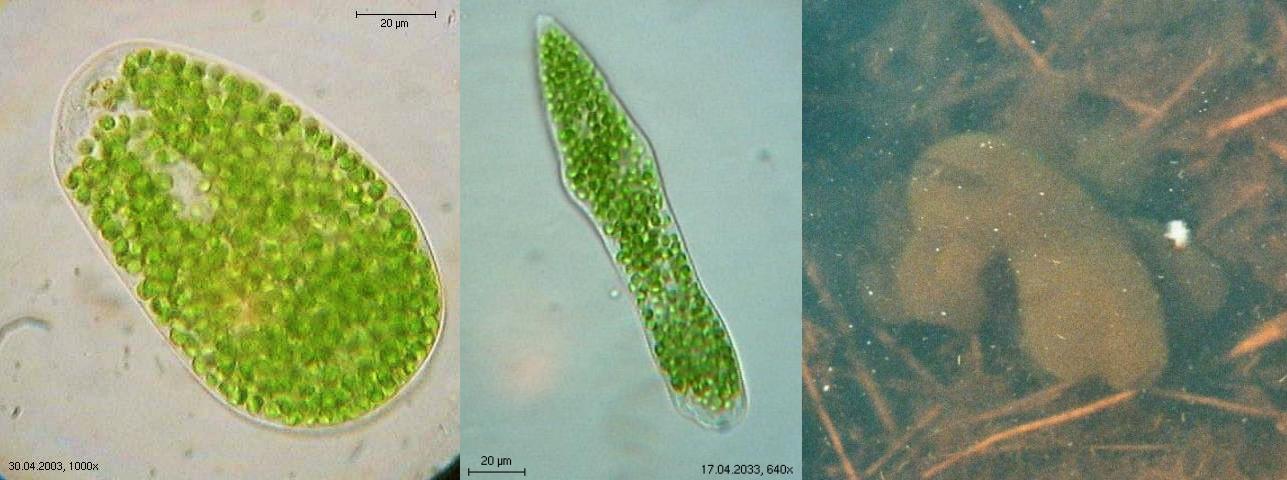
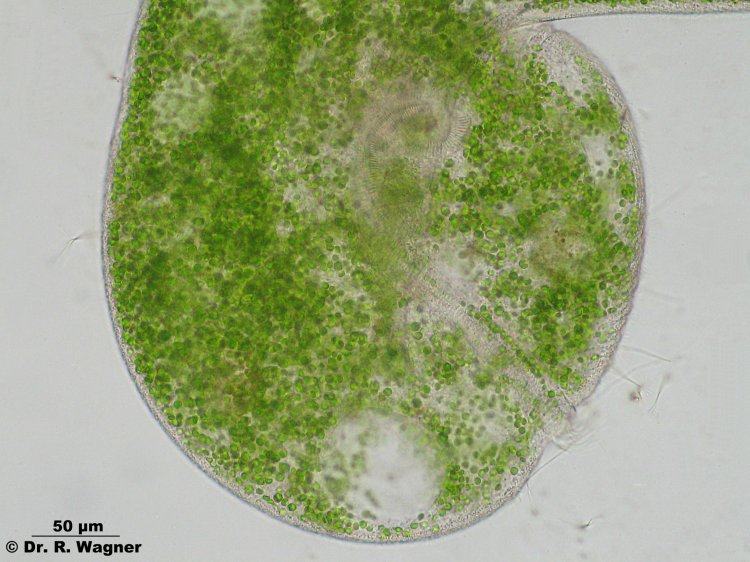
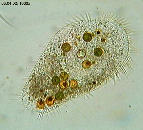
Frontonia spec. Galgenvenn, a heathland pond, June 2016
Shortly before discharging a food vacuoleMicroscopy, photomicrograph, Microfoto
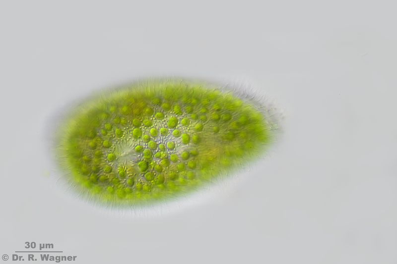
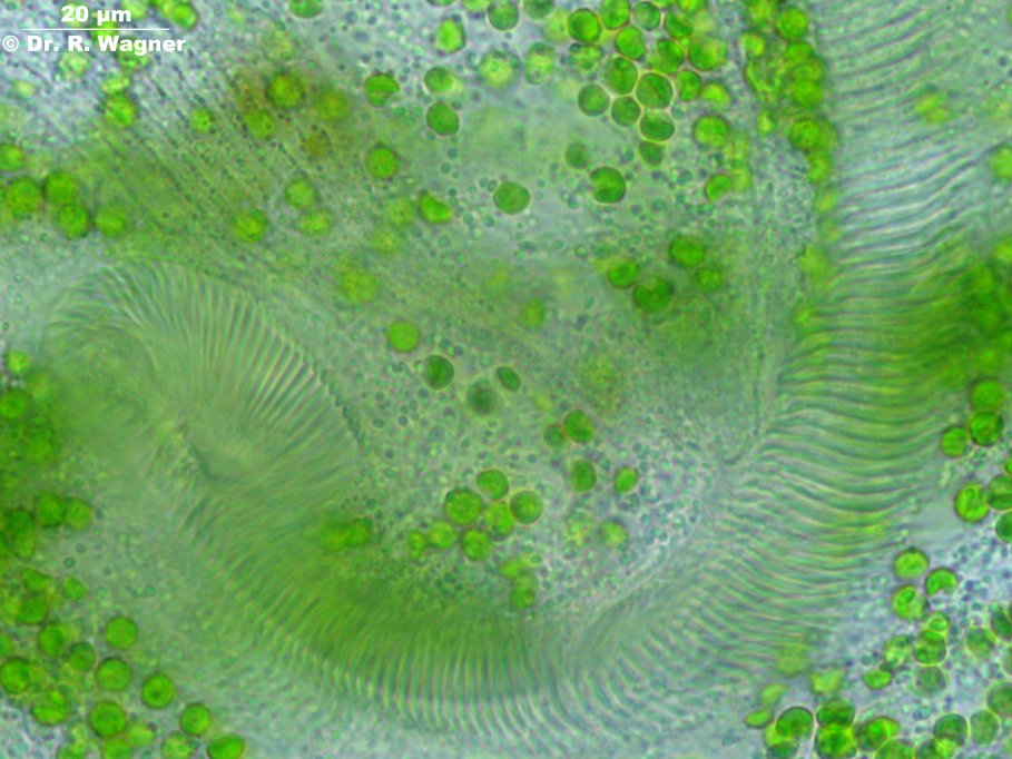
Frontonia atra botanical garden university Duesseldorf, April 2007
Special characteristic: the brownish granular in the plasma, which cotains symbiontic
ferro-bacteria, accumulating especially in the "head" - region.Microscopy, photomicrograph, Microfoto
Frontonia atra botanical garden university Duesseldorf, April 2007
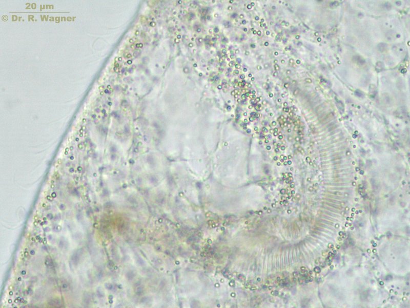
The contractile vacuole (CV) encircled by their feeding ducts and the food vacuole (FV)
are shown here Microscopy, photomicrograph, Microfoto
Frontonia atra botanical garden university Duesseldorf, April 2007
An obviously undigestible diatom is releasewd via a vacuole.Microscopy, photomicrograph, Microfoto
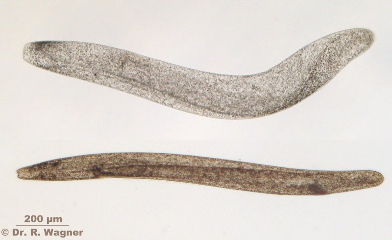
lacrymaria elegans putrid slime from my garden pond, September 2006Microscopy, photomicrograph, Microfoto
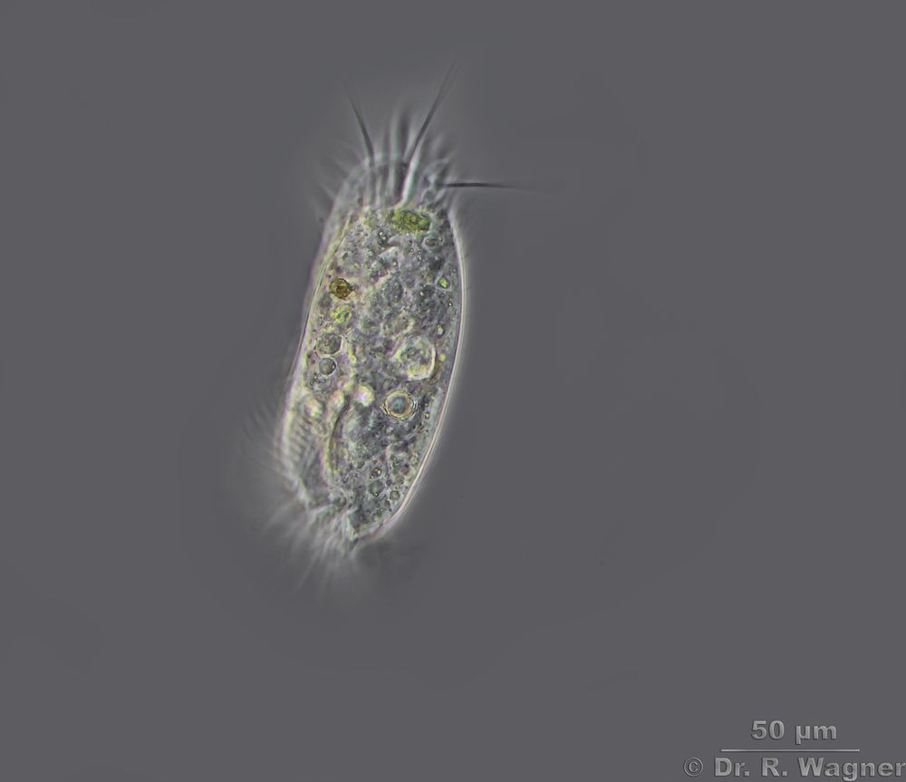
Litonotus cygnus garden pond, August 2020
Phasecontrast
Litonotus cygnus garden pond, August 2020
Phasecontrast
opercularia confusa national horticultural show Duesseldorf, spring 2001Microscopy, photomicrograph, Microfoto
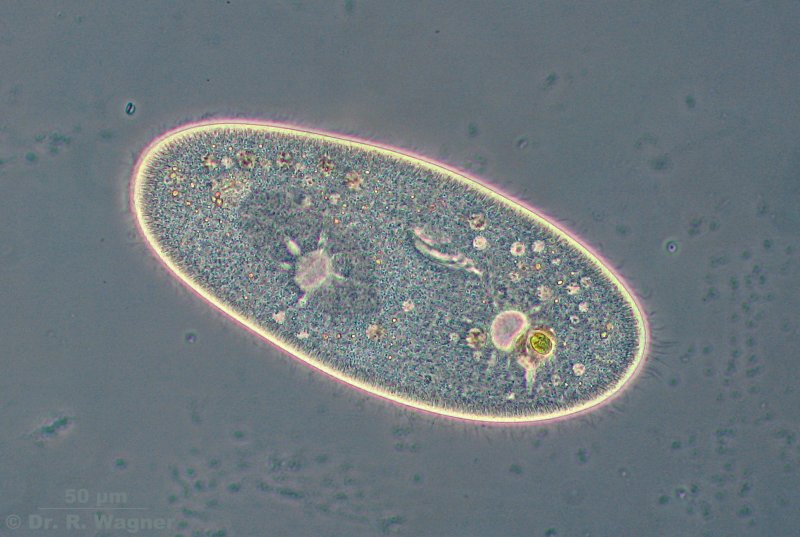
paramecium spec. garden pond, March 2007
This picture shows the 2 contractile vacuoles, the oral groove with the formation of a new
food vacuole at its end, and the overall surrounding cilia.Microscopy, photomicrograph, Microfoto
Paramecium spec. Gartenteich, März 2007
In the left part, a little bit hidden by the left contractile vacuole,
the macronucleus and close above a micronucleus
paramecium spec. garden pond, March 2007Microscopy, photomicrograph, Microfoto
end-phase of cell-division, only a small bridge is still connecting the two cells
Spirostomum ambiguum garden pond, April 2007
long, beads-like macronucleus, at the bottom a part of the also very long peristom,
lengthwise striped cell-wall with pigment-granulesMicroscopy, photomicrograph, Microfoto
Spirostomum ambiguum garden pond, April 2007Microscopy, photomicrograph, Microfoto
Stoma
Pseudovorticella monilata >garden pond, February 2007
Microscopy, photomicrograph, Microfoto
Stentor coeruleus Peringsmaar January 2022
Stentor coeruleus Peringsmaar January 2022
Stentor coeruleus Peringsmaar January 2022
Stentor coeruleus, phasecontrast Peringsmaar January 2022
stentor coeruleus botanical garden university Duesseldorf, February 2007
Microscopy, photomicrograph, Microfoto
stentor coeruleus botanical garden university Duesseldorf, February 2007
Microscopy, photomicrograph, Microfoto
Stentor igneus botanical garden university Duesseldorf, March 2007
stitch from 2 picturesMicroscopy, photomicrograph, Microfoto
Stentor igneus botanical garden university Duesseldorf, March 2007
Microscopy, photomicrograph, Microfoto
Stentor igneus botanical garden university Duesseldorf, March 2007
Microscopy, photomicrograph, Microfoto
stentor igneus botanical garden university Duesseldorf, November 2003
Overall view in phase contrast, The whole body surface is covered with cilia,
which are arranged in rows that correspond with striations on the cell body
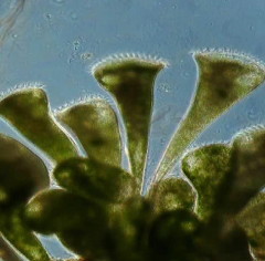
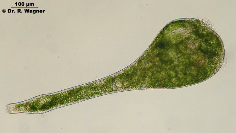
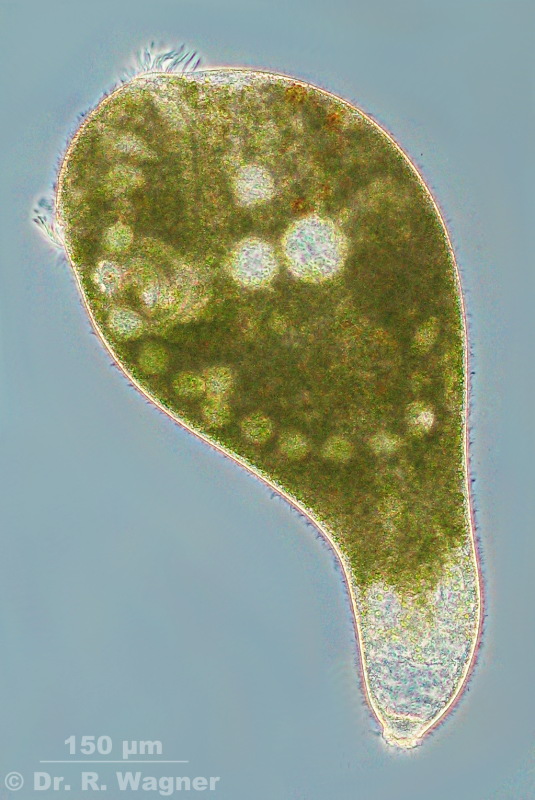
Stentor polymorphus garden pond, December 2007
Overall view in phase contrastMicroscopy, photomicrograph, Microfoto
Stentor polymorphus garden pond, December 2007
Overall view in phase contrastMicroscopy, photomicrograph, Microfoto
stentor polymorphus garden pond, dezember 2006
coordinated movement of the ciliaeMicroscopy, photomicrograph, Microfoto
stentor polymorphus garden pond, dezember 2006
The adoral zone of membranelles (AZM) encircles the thee peristome and winds into an
funnel-shaped part of the oral cavity that leads to the cytostome.
stentor polymorphus garden pond, dezember 2006
The adoral zone of membranelles (AZM) encircles the thee peristome and winds into an
funnel-shaped part of the oral cavity that leads to the cytostome.Microscopy, photomicrograph, Microfoto
stentor polymorphus garden pond, dezember 2006
The adoral zone of membranelles (AZM) encircles the thee peristome and winds into an
funnel-shaped part of the oral cavity that leads to the cytostome.
stentor polymorphus pond near the Wupper-dam, May 2006
probably a degenerated stentor polymorphus. besides the unusal form
the behavior is more similar to that of a dinoflagellat.
film, AVI-DIVX-format, 1,5 MBMicroscopy, photomicrograph, Microfoto
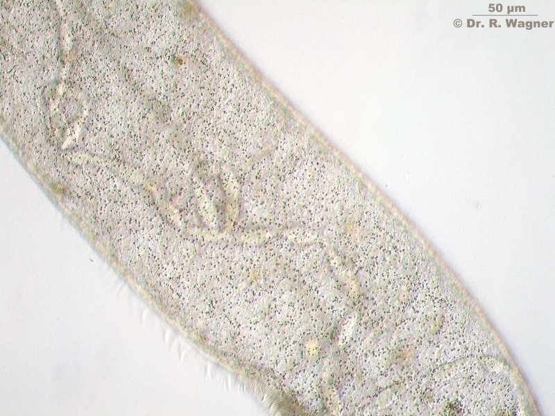
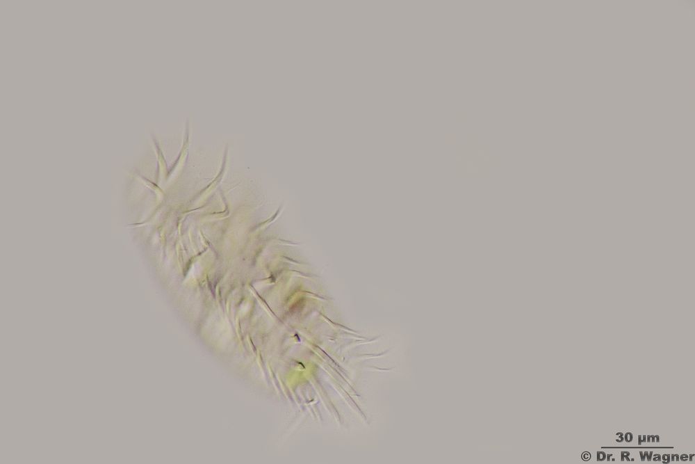
stichotricha secunda pond near the Wupper-dam, May 2006
Microscopy, photomicrograph, Microfoto
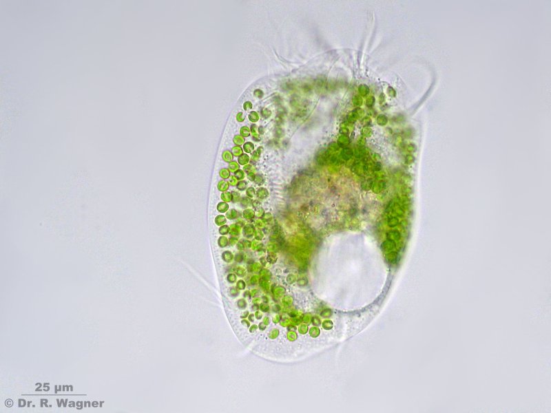
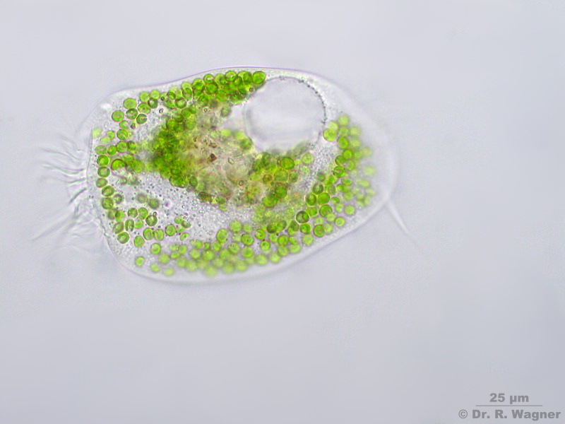
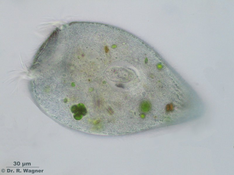
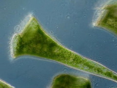
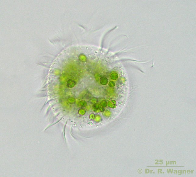
Strombidium viride garden pond March 2007
The green algae in the plasma are not symbiontic zoochlorellas, but intact plastids of eaten algae
Strombidium viride garden pond March 2007
The green algae in the plasma are not symbiontic zoochlorellas, but intact plastids of eaten algae
stylonychia garden pond, April 2002
Microscopy, photomicrograph, Microfoto
stylonychia mytilus botanical garden university Duesseldorf,, August 2006
Microscopy, photomicrograph, Microfoto
Stylonychia old clay pit, May 2015
phasecontrast
Stylonychia old clay pit, May 2015
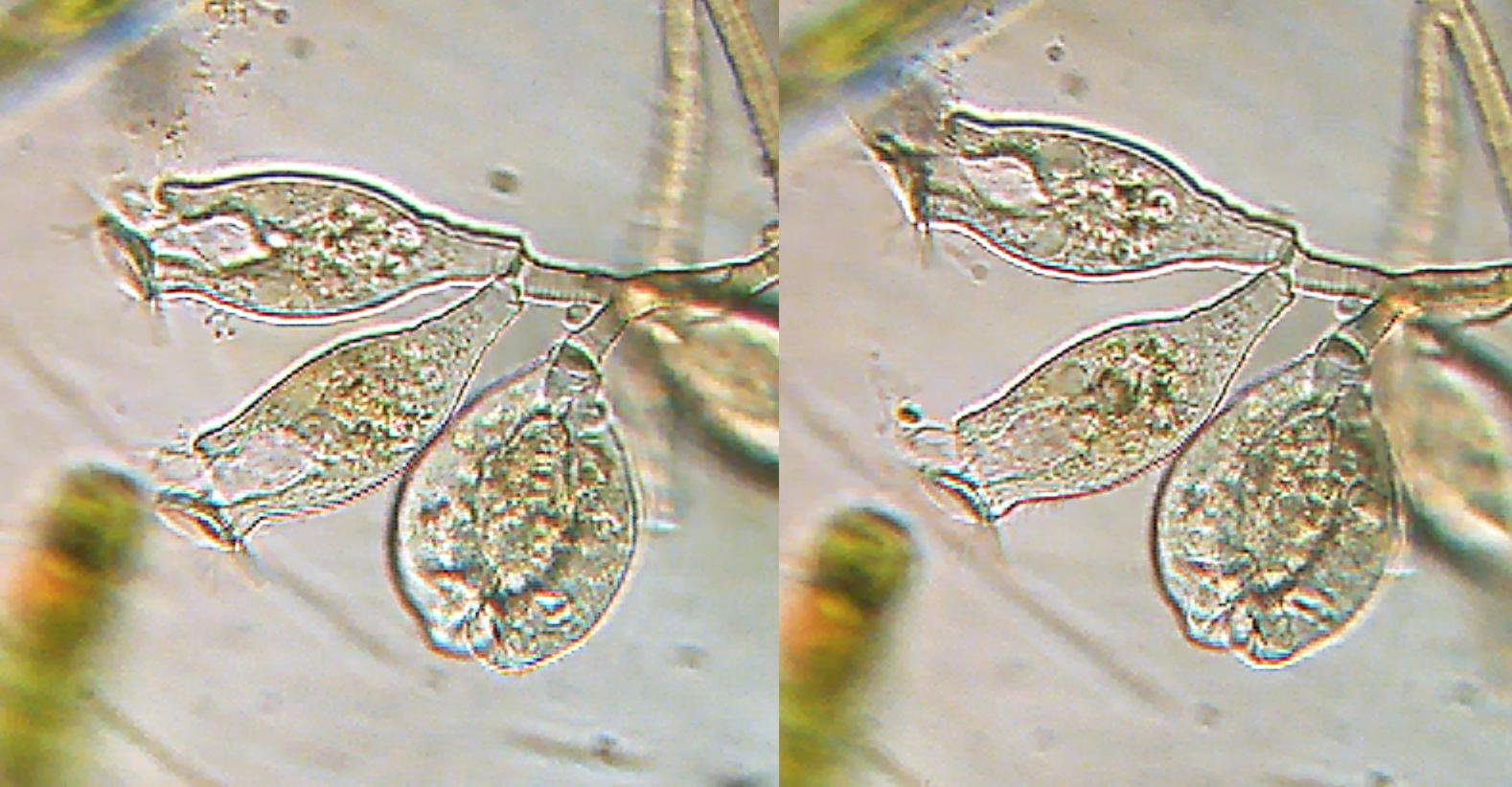
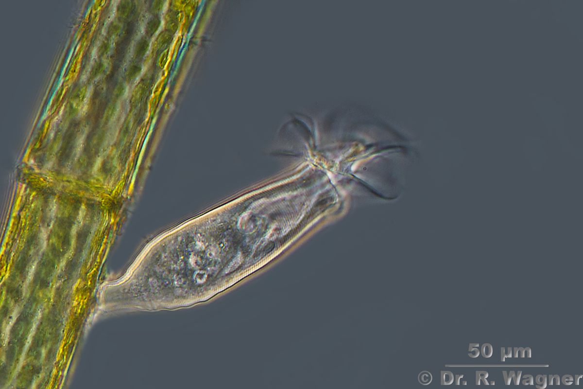
Thuricola folliculata national horticultural show Duesseldorf, March 2007
2 animals in one lorica, here the seldomm form without green zoochlorellas. Characteric
is the valve that closes the lorica when the animals have drwan back.Microscopy, photomicrograph, Microfoto
Thuricola folliculata national horticultural show Duesseldorf, March 2007
2 animals in one lorica, here the seldomm form without green zoochlorellas. Characteric
is the valve that closes the lorica when the animals have drwan back.
Thuricola folliculata national horticultural show Duesseldorf, March 2007
2 animals in one lorica, here the seldomm form without green zoochlorellas. Characteric
is the valve that closes the lorica when the animals have drwan back.Microscopy, photomicrograph, Microfoto
Thuricola folliculata national horticultural show Duesseldorf, March 2007
Video (DIVX-format, ~3 MB)Microscopy, photomicrograph, Microfoto
Thuricola folliculata Small Pond near Hilden, May 2011
YouTube video
Thuricola folliculata Small Pond near Hilden, May 2011
Microscopy, photomicrograph, Microfoto
Thuricola folliculata national horticultural show Duesseldorf, March 2007
2 animals in one loricaMicroscopy, photomicrograph, Microfoto
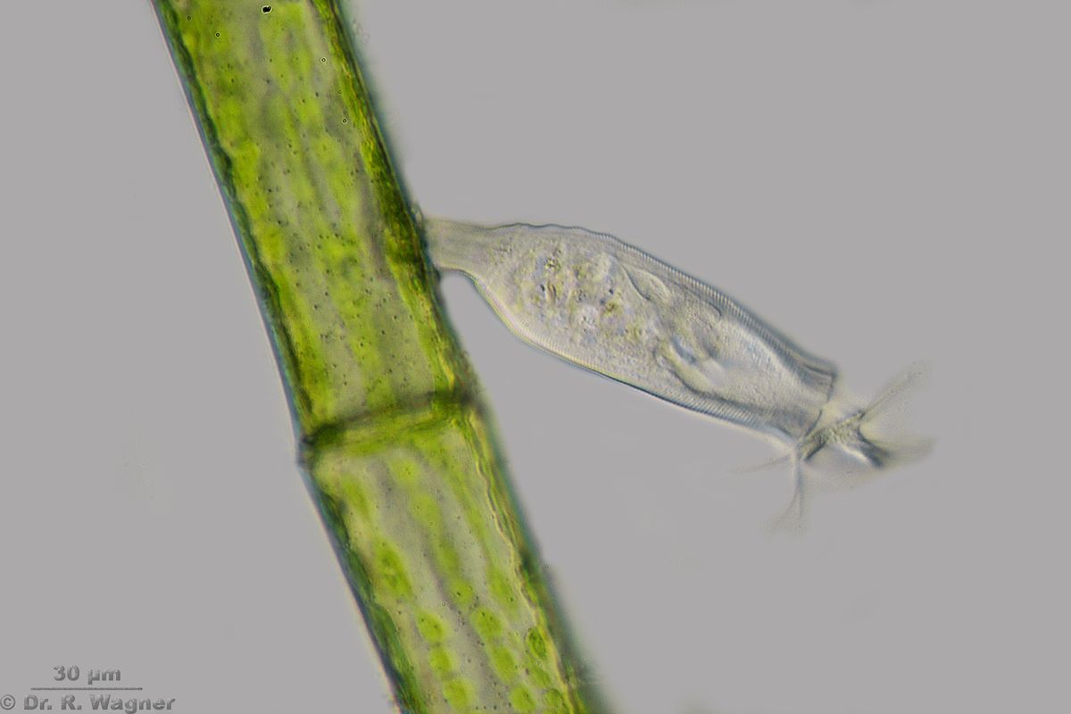
Thuricola folliculata national horticultural show Duesseldorf, March 2007
2 animals in one lorica, living in symbiosis with green zoochlorellas
thuricola folliculata national horticultural show Duesseldorf, March 2007
video, AVI-DIVX-format, 2,9 MB
thuricola folliculata national horticultural show Duesseldorf, June 2006
Microscopy, photomicrograph, Microfoto
thuricola folliculata garden pond, April 2006
video, AVI-DIVX-Format, 1,5 MB
vaginicola botanical garden university Duesseldorf, spring 2001
Microscopy, photomicrograph, Microfoto
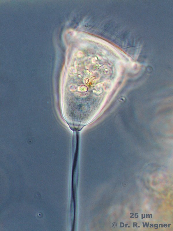
Vorticella spec. garden pond April 2007
Sometimes Vorticella contracts its stem to a helix. This is done with the aid of the myonem-bundle inside the stem
In this phase-contrast picture you can the myonem-bundle inside the stem and even how it divides into two
different lines when entering the cell-body.
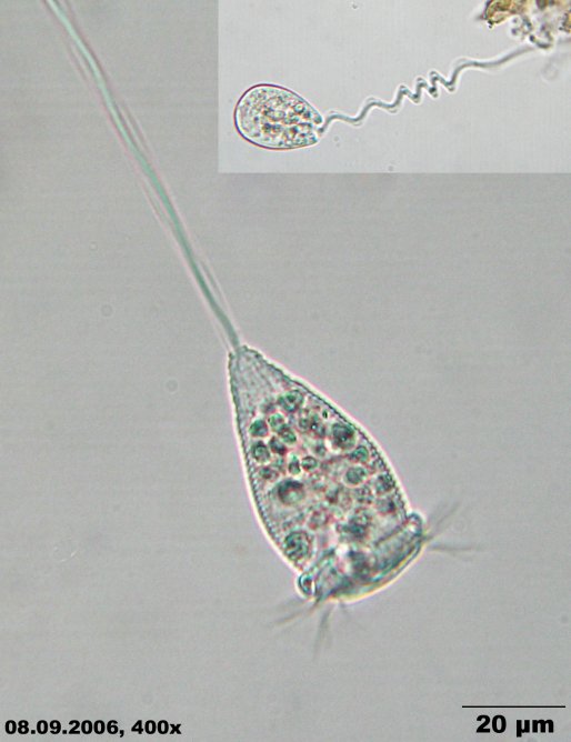
vorticella microstoma garden pond, September 2006Microscopy, photomicrograph, Microfoto
Vorticella spec. Phasecontrast, Garden pond March 2017
YouTube video
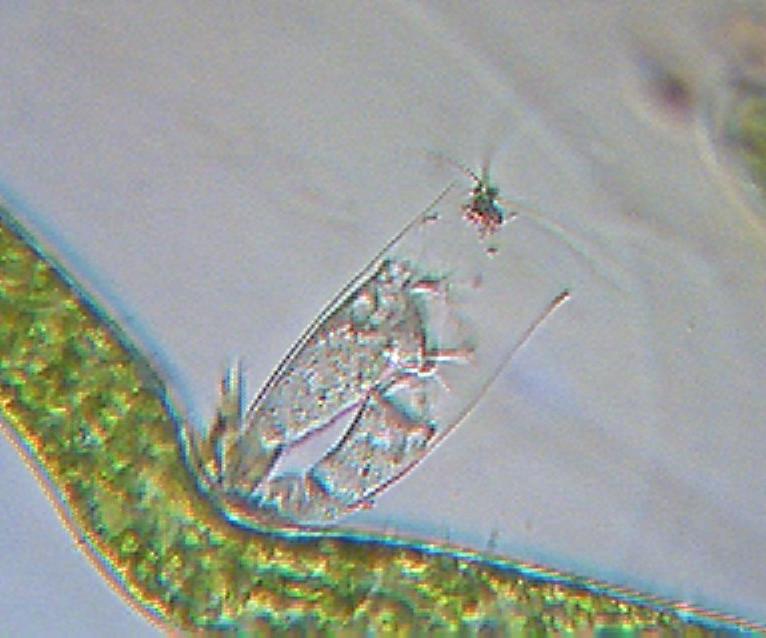
vorticella spec. national horticultural show Duesseldorf, June 2006
Ein Glockentierchen bei der Arbeit
video, wmv-format, 1,6 MB